X-ray and dental tomography laboratory

Dental X-ray Opole
and dental tomography

Modern dental diagnostics in Opole

ADENTA X-ray and dental tomography laboratory

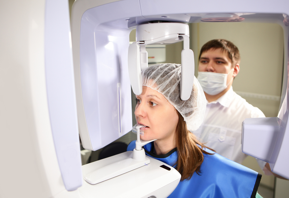
Our ADENTA dental clinic has a modern digital X-ray laboratory. It is equipped with a versatile diagnostic tool for CBCT (Con), pantomographic and cephalometric imaging, a modern (developed in 2023) Dexis OP 3D device. It is characterized by a very low radiation dose, and taking a panoramic picture takes only 9 seconds. Four different 3D imaging fields allow them to be adapted to individual needs. High-class equipment for performing extra- and intraoral X-ray examinations allows us to fully determine the patient's needs and accurately plan the directions of treatment.

We perform examinations for our patients and on behalf of other facilities:

  • Intraoral spot X-rays of single teeth
  • Pantomographic overview images of the entire dentition - maxilla and mandible, including pediatric and partial pantomographic and temporomandibular joints
  • Cephalometric and PA, including pediatric
  • Computed tomography (CBCT) of the full maxilla and mandible, temporomandibular joints, sinuses and individual teeth
Examinations for our patients and on behalf of other facilities

Take advantage of modern dental diagnostics at the Adenta Clinic.

Comprehensive treatment and all necessary examinations in one place!

Intraoral spot X-rays

  1. Intraoral spot X-rays Small spot X-rays are the basic radiological images during dental treatment. On these images, we see hard structures - teeth and bones. Due to the different absorption of X-rays by tissues, we can see deviations from the normal X-ray image, through which we detect pathological changes. These images are widely used in conservative dentistry, endodontics and for implantological control. It is not uncommon for a spot X-ray of a tooth to be taken multiple times. This is related to the radiological control of individual stages of treatment.
  2. Pantomographic overview images of the entire dentition Panoramic images, also known as pantomographic or OPG, are the first and basic method of assessing the condition of: teeth, bones, soft tissues and joints in the facial area of the skull. It should be remembered that this is an overview image, which should be supplemented with more accurate X-ray imaging, such as a spot X-ray or CBCT. We most often recommend a pantomogram:
    • for a general analysis of the state of the dentition, the presence of tooth buds, impacted teeth, e.g. third molars,
    • for injuries to the maxilla, mandible or teeth,
    • for the analysis of the state of hard tissues - the state of the periodontium, the presence of pathological changes in the jaw bones,
    • if sialolithiasis is suspected,
    • during an ongoing inflammation or infection,
    • for planning treatment: implantological, prosthetic, orthodontic,
    • before a planned tooth extraction,
    • in the diagnosis of the presence of pathological changes in the oral cavity.
    • in case of problems with the temporomandibular joints.
  3. Cephalometric X-ray Cephalometric X-rays (lateral skull distance images) are necessary for measurements and planning of orthodontic treatment, and for the assessment of possible facial asymmetries, it is also possible to perform an examination in the PA (postero-anterior) projection.
  4. Computed tomography Dental computed tomography - CBCT, i.e. a three-dimensional image of soft and hard tissues with a layer accuracy of a fraction of a millimeter!!
Precise diagnostics with the Dexis OP 3D device

Precise diagnostics with the Dexis OP 3D device

The imaging fields of our Dexis OP 3D device have been selected based on actual clinical needs:

  • 5x5 - the highest quality image is taken during endodontic treatment. The layer thickness is the smallest and allows the endodontist to accurately visualize the root canals, their course and position. Changes at the apex of the tooth roots, fractures or cracks of the tooth or possibly tools in the canal are very well visible. Such a small imaging field can also be useful for a surgeon.
  • 6x9 - is great for controlling the maxilla or mandible in planning implant-prosthetic treatment of a smaller range up to several points.
  • 9x11 - planning treatment in two dental arches - upper and lower at the same time. A very good projection for the surgeon for possible extractions of all wisdom teeth.
  • 9x14 - the largest imaging field allows you to see the temporomandibular joints and airways. This is particularly useful for patients with sleep apnea and for planning and monitoring orthodontic or orthognathic treatment.

Diagnostics in various fields of dentistry

Application of individual types of radiological examinations in various fields of dentistry:

X-ray laboratory at the Adenta Clinic

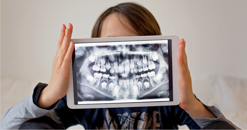
At our Adenta Clinic, we perform each of the above-mentioned projections in a separate room of the X-ray laboratory on the latest equipment with the use of the lowest possible radiation dose. The examination is visible in real time on the computer monitor and in the office. Comprehensive and accurate X-ray diagnostics along with an interview is the beginning of the entire dental treatment process. It allows for a full assessment of the condition of the teeth, bones, gums, temporomandibular joints, as well as the paranasal sinuses and upper respiratory tract in order to create an individualized comprehensive treatment plan. It is also used to control the course of treatment and assess its quality.

We also perform imaging diagnostics on behalf of other facilities. The examination is downloaded from the "cloud" and sent by e-mail with data protection.

Examinations for our patients and on behalf of other facilities

Opinions of our patients

Star Rating
Dr. Asia is not only very nice but also fully professional in her approach to the patient. Me and my teeth highly recommend her :)
Kacha Gie
Star Rating
Professionalism in every way! The doctor is always helpful with advice, I recommend! :-)
Żaneta Górna
Star Rating
The Doctor's approach is very professional, I recommend him to everyone!!! My fear of treatment was overcome thanks to the kind, reliable, and friendly service, and the tooth problem was solved. :)
Natalia Kulon
Star Rating
Dr. Joasia is a professional dentist!!! Warm, fully committed, and passionate! I recommend 100%.
Emilia Jończyk-Gorzelska
Star Rating
Highly recommend!! Professional approach to the patient and a very friendly team, what more could you ask for!
Karolina Przybyła
Star Rating
Highly recommend! Full professionalism and commitment! I use the services regularly :-)
Łukasz Radziszewski
Star Rating
The doctor is a professional in every respect. He always gives good advice. The team is well-coordinated. I highly recommend him, and so do my teeth.
Katarzyna Rymarz
Star Rating
Fantastic dentist! Gentle, professional and personal. I highly recommend Adenta!
Patrizia Isabelle Duda
Star Rating
I highly recommend. Full professionalism in service execution and a wonderful approach to the patient. The best dentist in Opole.
Maya Madeline

Get a beautiful, healthy smile - without pain and stress

Call and make an appointment!

Banner MediRaty